

Interactive CT & MRI Anat.Lite

توضیحات Interactive CT & MRI Anat.Lite
★Lite version★
This is the free Lite version of "Interactive CT and MRI Anatomy".
The function is restricted.
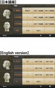
You can only see the transverse CT images of the head.
Please check the operation before purchasing the full version.
★ Details ★
This application is developed for medical students, interns, residents, doctors, nurses, and radiology technicians to understand the essential anatomical terms of the body.
You can learn anatomy by answering the terms by step-to-step questions using a total of 241 CT and MRI images.
A total of 17 images of 3D-CT, MRA and plain X-ray film(particularly the extremities) are included as references.
Other reference images include coronary artery segments defined by the American Heart Association(AHA), pulmonary segments, and liver segments(according to Couinaud classification).
You can enlarge all the images by simple manipulation.
★ Major functions ★
There are 4 major functions.
-1) Anatomical mode
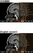
Anatomical terms are overlaid on the images.
It can be used as the anatomical atlas.
-2) Quiz mode type 1
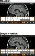
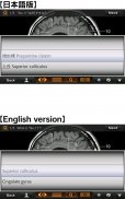
You select the part of the image by using anatomical term.
Questions will basically appear randomly.
-3) Quiz mode type 2
You select the anatomical term by the part of the image.
Questions will basically appear randomly.
-4) Index
You can find the specific images by using anatomical terms.
★ Intended users ★
-Medical students
-Interns and residents
-Doctrors
-Nurses
-Radiology technicians
-All those who are intrested in CT and MRI anatomy
★ Images(a total of 258 images) ★
Images basically include horizontal, coronal, and sagital planes.
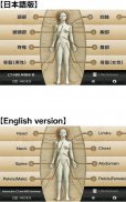
-Head(36 images including CTA and 3D-CT)
-Neck(24 images)
-Spine(19 images including plain X-ray films)
-Chest(61 images including 3D-CT images)
-Abdomen (37 images)
-Pelves: male (9 images)
-Pelvis: female (11 images)
-Extremities (shoulder, hand, elbow, hip joint, knee, foot) (61 images including plain X-ray films)
Editors
Toshiaki Nitori, M.D. (Professor of Radiology, Kyorin University, School of Medicine)
Yasuo Sasaki, M.D. (Manager of diagnostic radiology, Iwate Prefectural Central Hospital)
</div> <div jsname="WJz9Hc" style="display:none">★ Lite-versie ★
Dit is de gratis Lite-versie van "Interactive CT en MRI Anatomie".
De functie is beperkt.
U kunt alleen de dwarse CT-beelden van het hoofd te zien.
Controleer de werking vóór de aankoop van de volledige versie.
★ Details ★
Deze applicatie is ontwikkeld voor medische studenten, stagiaires, bewoners, artsen, verpleegkundigen en radiologie technici om de essentiële anatomische voorwaarden van het lichaam te begrijpen.
U kunt anatomie leren door het beantwoorden van de voorwaarden voor stap-voor-stap vragen met behulp van een totaal van 241 CT en MRI-beelden.
Een totaal van 17 beelden van de 3D-CT, MRA en plain X-ray film (met name de extremiteiten) worden opgenomen als gevonden.
Andere referentiebeelden omvatten coronaire segmenten gedefinieerd door de American Heart Association (AHA), pulmonaire segmenten en segmenten lever (volgens Couinaud classificatie).
U kunt vergroten alle beelden door een eenvoudige manipulatie.
★ Belangrijke functies ★
Er zijn 4 belangrijke functies.
-1) Anatomische modus
Anatomische termen worden overlay op de beelden.
Het kan worden gebruikt als anatomische atlas.
-2) Quiz modus type 1
U selecteert het deel van het beeld met behulp van anatomische termijn.
Vragen zal in principe verschijnen willekeurig.
-3) Quiz mode type 2
U selecteert de anatomische term door het deel van het beeld.
Vragen zal in principe verschijnen willekeurig.
-4) Index
U kunt de specifieke beelden met behulp van anatomische termen te vinden.
★ Beoogde gebruikers ★
-Medische Studenten
-Interns En bewoners
-Doctrors
-Verpleegkundigen
-Radiology Technici
-alle Degenen die geintresseerd in CT en MRI anatomie
★ Afbeeldingen (in totaal 258 foto's) ★
Afbeeldingen in principe onder horizontaal, coronale en sagittale vliegtuigen.
-hoofd (36 beelden inclusief CTA en 3D-CT)
-Neck (24 foto's)
-Spine (19 beelden inclusief plain X-ray-films)
-Chest (61 beelden inclusief 3D-CT-beelden)
-Abdomen (37 foto's)
-Pelves: Mannelijk (9 beelden)
-Pelvis: Vrouw (11 foto's)
-Extremities (Schouder, hand, elleboog, heup, knie, voet) (61 beelden inclusief plain X-ray-films)
Editors
Toshiaki Nitori, MD (hoogleraar Radiologie, Kyorin University, School of Medicine)
Yasuo Sasaki, MD (Manager van radiodiagnostiek, Iwate Prefectural Central Hospital)</div> <div class="show-more-end">


